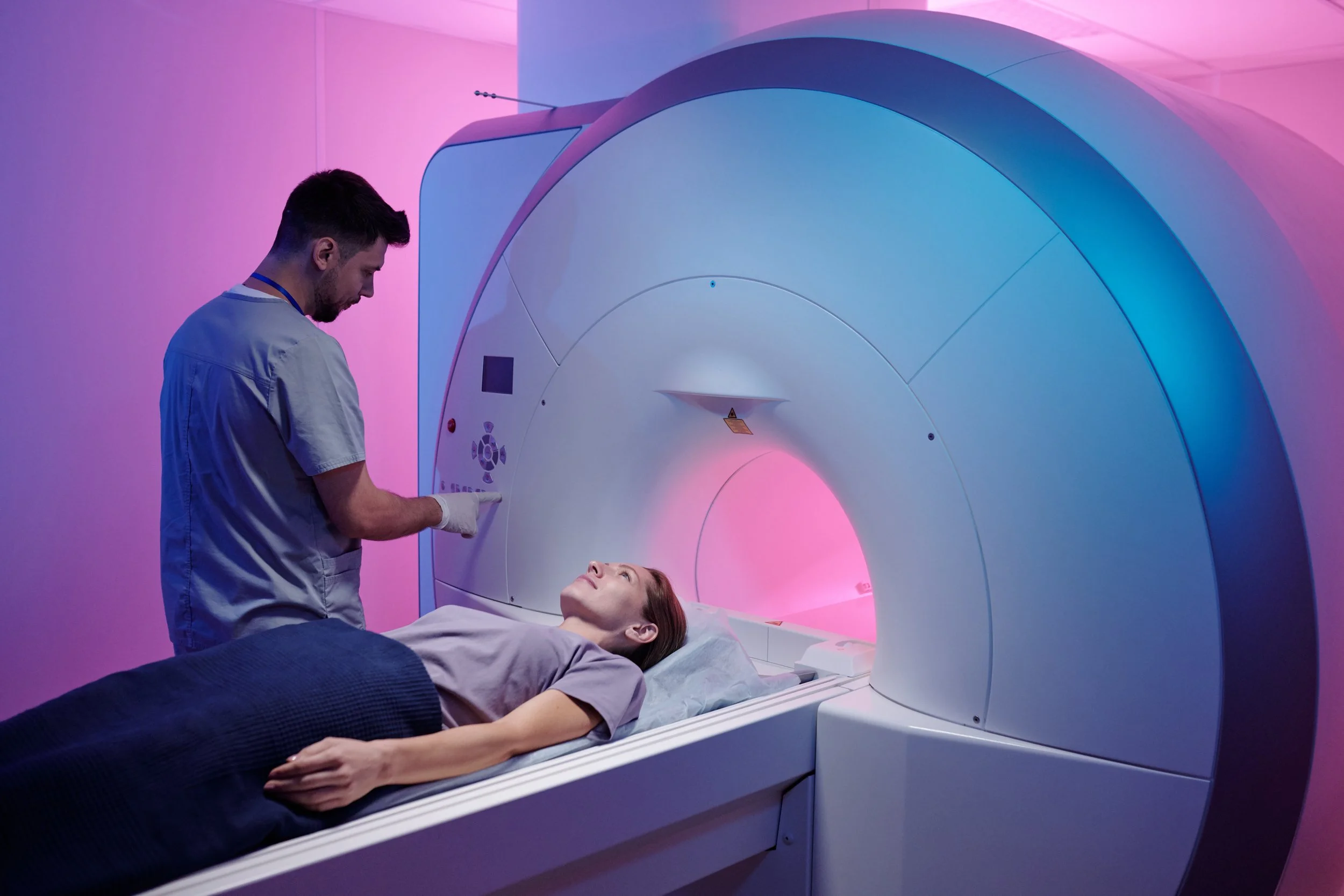
Elite Sport: AI Muscle Analytics
Launching the AI-based technology across our imaging network. The technology takes segments and analyzes musculoskeletal (MSK) data for comprehensive muscle health analysis.
The software precisely quantifies individual muscle volume and quality, fat fraction, left-right asymmetries, as well as scar tissue, edema, tendon and bone morphology to drive better health and performance outcomes.
We have teamed up with Springbok Analytics to bring their software to some of our imaging sites to allow our teams to understand their players muscle health.
Springbok’s AI-based technology segments and analyzes musculoskeletal (MSK) data for comprehensive muscle health analysis.
Springbok precisely quantifies individual muscle volume and quality, fat fraction, left-right asymmetries, as well as scar tissue, edema, tendon and bone morphology to drive better health and performance outcomes.
Initial Locations
-
Prime Health, London.
The service will be available when we go live at Prime Health, Queen Anne Street, London.
Appointments bookable online.
-
Prime Health, Weybridge.
The service will be available when we go live at Prime Health, Weybridge.
Appointments bookable online.
-
Medical Imaging Partnership, Stockport.
The service will be available when we go live at Medical Imaging Partnership, Stockport, Manchester.
Appointments bookable online.
Wish to discuss this further?
This service is coming soon. If you wish to be added to our waiting list for contact and launch complete the form opposite.
Thank you.
Further Information
-
Updated: Apr 23
Understanding an athlete’s body composition has long been a cornerstone of sports science and medicine. Integrating anthropometry and adiposity assessments is standard practice. While clearly crucial in weight classification sports, quantifying body composition is valuable across various sports. It enables practitioners to evaluate an athlete’s physical condition and monitor changes over a season in response to training programmes, nutritional interventions, or injuries, among other factors.
However, there is a compelling case for expanding our definition of ‘body composition’ to include a comprehensive understanding of musculoskeletal health. This broader perspective supports various applications across health, performance, and injury.
This article explores the evolving landscape of body composition analysis, highlighting both traditional methods and the supplementary insights provided by technological advancements. We will particularly focus on the development of AI-driven MRI analysis by Springbok Analytics, which offers detailed insight into the musculoskeletal health of athletes, and how it differs from other technologies.
Body Composition Analysis: A Brief Overview
Traditionally, body composition assessments focus on body mass, lean mass, fat mass, and where possible, bone mineral content. These assessments are particularly relevant in sports where athletes resist gravity, such as when jumping and running, where excess fat mass is perceived as ‘dead weight’ (Kasper et al., 2021).
Options for body composition evaluation range from rudimental measurements to increasingly advanced technology (for a comprehensive, open access review see Kasper et al., (2021)). Simple assessments of physique, such as Body Mass Index (BMI) and the waist-to-hip ratio, provide an initial understanding and are especially useful in public health settings.
The next level utilises techniques that estimate fat and fat-free mass, including skinfold callipers, hydrostatic weighing (densitometry), air displacement plethysmography (commonly recognised by its commercial name: BOD POD), and bioelectrical impedance analysis.
Finally, newer technologies offer deeper insights into body composition. Dual-energy X-ray absorptiometry (DXA or DEXA) discerns bone and soft tissue using X-rays. Computed tomography (CT) provides tissue volume measurements from high-resolution X-ray images, and magnetic resonance imaging (MRI) distinguishes soft tissues based on different magnetic properties of cells.
Springbok Analytics: The AI-Driven Evolution in MRI Technology
Unlike DXA and CT, which use X-rays and thus involve exposure to ionising radiation, MRI scans utilise magnetic properties to comprehensively examine both soft and hard tissues. This allows for analysis beyond adiposity and bone, by evaluating muscle and tendon morphology. While traditional analysis was marred by time-intensive manual segmentation of MRI ‘slices’, machine learning has significantly reduced these extensive processing times (Ni et al., 2019).
Springbok Analytics harnesses AI to automate MRI scan analysis, creating a ‘digital twin’ – a digital representation of an individual's musculature. This 3D model enables calculation of:
Muscle volume
Left to right asymmetries
Muscle fat infiltration
Tendon morphology in the hamstring muscle
Aponeurosis cross sectional areas and area of attachment with muscle
Scar tissue and edema volume
Traditionally, imaging focuses on a specific region of musculoskeletal tissue (e.g., “They have a hamstring issue, so that region will be scanned”). Now, broader capture of full body regions offers a more comprehensive understanding of musculoskeletal health in our athletes. A single lower extremity MRI scan allows Springbok to analyse all 76 muscles and 12 skeletal structures, namely both left and right femurs, patellas, tibias, fibulas, pelvis, as well as the sacrum and vertebrae. Measures can be tracked over time for an individual and compared against a comprehensive normative database using the Springbok Score.
This information can expand the understanding of an athlete’s physical condition. It allows practitioners to profile an athlete’s musculoskeletal health, observe shifts in response to nutritional intervention, quantify asymmetries between the left and right sides that could heighten injury risk, and identify the magnitude and location of muscle changes due to a training programme.
This exciting technology exemplifies how AI can potentially enhance our understanding of athlete health and management. So where might this fit within our current sports performance support processes?
Expanding our Definition of ‘Body Composition’
Access to DXA scans has increased in professional sports in recent times, though often limited in certain settings by budget constraints. Yet, DXA and the MRI analysis detailed above should not be confused. They are different, but potentially complementary technologies.
DXA, a two-dimensional imaging technique, uses X-rays at two energy levels to separate images into bone and soft tissue components. It is the 'gold standard' for bone mineral density measurement and assesses total and regional adiposity. However, research suggests it may not consistently evaluate muscle tissue changes over time (Tavoian et al., 2019).
This is where Springbok’s AI-driven analytics on MRI scans adds further depth of understanding. We’ve previously discussed on the blog the importance of changing perspectives; of intentionally zooming in and out to adjust your viewpoint. This technology takes the ‘zoom button’ even further, offering a detailed approach to musculoskeletal health. This understanding complements "traditional" body composition measures, whether through DXA, skinfolds, etc., by providing a broader comprehension of 'body composition'.
Applications in Injury Rehabilitation and Beyond
MRI scans are often first thought of in relation to injury, and there is certainly significant interest in using this technology in this area. Springbok’s Learning Center include case studies of how this analysis has aided athletes returning from injury, such as a professional athlete post ACL-reconstruction, a collegiate football (soccer) player post-ankle ligament reconstruction, and a collegiate lacrosse athlete with chronic knee pain.
Springbok Analytics Case Study: Targeted ACL Rehabilitation Programme
However, prevention is better than cure. A more granular understanding of musculoskeletal health may help mitigate injury risk. Springbok’s patented MRI analysis is part of a 3-year prospective study in NCAA Division I collegiate [American] football players (Heiderscheit et al., 2022). Approximately 560 student-athletes will be assessed via measures of strength, sprinting biomechanics, and Springbok Analytics MRI examinations, as the group seek to develop a hamstring injury index (HAMIR).
There are also opportunities to explore this data from a profiling and performance perspective, particularly in athletes from asymmetrical sports. These athletes adapt to their sport's demands, often developing asymmetries. While some asymmetries may be performance-driven or even advantageous, there is a tipping point. It is challenging to determine the magnitude of asymmetry that might increase injury risk. Understanding and tracking morphological asymmetries over time may provide more objective and detailed information to align with performance and injury issues.
Final Thoughts
This emerging technology allows us to explore the question, ‘How is the athletic body composed?’ with greater detail and precision. While traditional methods of body composition analysis provide valuable insight into anthropometry, adiposity, and bone density, they can now be complemented by a more detailed quantification of musculoskeletal health.
Gaining a comprehensive and nuanced understanding of an athlete’s body from a muscular perspective can further our efforts to mitigate injury risk, facilitate return from injury, and tailor athlete profiling to the specific demands of their sport. As with any potential technological implementation, careful consideration of how it will translate into a particular setting is necessary. This includes evaluating its value against the potential burden. Ultimately, it may be time to broaden our definition of ‘body composition’ and consider how a more encompassing view of muscle health can enhance our support for athletes.
-
Purpose
Human skeletal muscle has the profound ability to hypertrophy in response to resistance training (RT). However, this has a high energy and protein cost and is presumably mainly restricted to recruited muscles. It remains largely unknown what happens with nonrecruited muscles during RT. This study investigated the volume changes of 17 recruited and 13 nonrecruited muscles during a 10-wk single-joint RT program targeting upper arm and upper leg musculature.
Methods
Muscle volume changes were measured by manual or automatic 3D segmentation in 21 RT novices. Subjects ate ad libitum during the study and energy and protein intake were assessed by self-reported diaries.
Results
Posttraining, all recruited muscles increased in volume (range: +2.2% to +17.7%, P < 0.05), whereas the nonrecruited adductor magnus (mean: −1.5% ± 3.1%, P = 0.038) and soleus (−2.4% ± 2.3%, P = 0.0004) decreased in volume. Net muscle growth (r = 0.453, P = 0.045) and changes in adductor magnus volume (r = 0.450, P = 0.047) were positively associated with protein intake. Changes in total nonrecruited muscle volume (r = 0.469, P = 0.037), adductor magnus (r = 0.640, P = 0.002), adductor longus (r = 0.465, P = 0.039), and soleus muscle volume (r = 0.481, P = 0.032) were positively related to energy intake. When subjects were divided into a HIGH or LOW energy intake group, overall nonrecruited muscle volume (−1.7% ± 2.0%), adductor longus (−5.6% ± 3.7%), adductor magnus (−2.8% ± 2.4%), and soleus volume (−3.7% ± 1.8%) decreased significantly (P < 0.05) in the LOW but not the HIGH group.
Conclusions
To our knowledge, this is the first study documenting that some non recruited muscles significantly atrophy during a period of RT. Our data therefore suggest muscle mass reallocation, that is, that hypertrophy in recruited muscles takes place at the expense of atrophy in non recruited muscles, especially when energy and protein availability are limited.


